Everything You Need To Know Related To Pain In The Arches
Overview
Posterior tibial tendon dysfunction is one of several terms to describe a painful, progressive flatfoot deformity in adults. Other terms include posterior tibial tendon insufficiency and adult acquired flatfoot. The term adult acquired flatfoot is more appropriate because it allows a broader recognition of causative factors, not only limited to the posterior tibial tendon, an event where the posterior tibial tendon looses strength and function. The adult acquired flatfoot is a progressive, symptomatic (painful) deformity resulting from gradual stretch (attenuation) of the tibialis posterior tendon as well as the ligaments that support the arch of the foot.
Causes
Foot cramps are caused by muscles suddenly spasming uncontrollably. They most commonly cause foot arch pain but can occur anywhere in the foot and lower leg. Usually, they only last a few seconds but in more extreme cases they can continue longer. Often, there is no obvious reason why people suffer from foot cramps, but possible causes include diet, muscle tightness and weakness, dehydration, reduced circulation and fatigue. Sometimes, it can be a sign of an underlying medical condition so if the problem keeps recurring, do consult your doctor. Some of the best ways to reduce the incidence of foot arch pain from cramps include doing exercises, using heat, drinking plenty of water, using toe stretchers and ensuring you are wearing good footwear.
Symptoms
Bones and ligaments work together to form joints, and bones are joined together by ligaments. Strains occur in ligaments. In the arch, there are ligaments that are located at the ends of each bone. These ligaments connect the bones to other bones on both ends and on the sides. Point tenderness and looseness of a joint are indicators of a sprain. Fractures are indicated by point tenderness that may be severe over the area of bone that is affected. There may be a distinguishable lump or gap at the site of the fracture. A rotated toe or forefoot may also be a sign of a fracture.
Diagnosis
After you describe your symptoms and discuss your concerns, your doctor will examine your foot. Your doctor will look for these signs. A high arch. An area of maximum tenderness on the bottom of your foot, just in front of your heel bone. Pain that gets worse when you flex your foot and the doctor pushes on the plantar fascia. The pain improves when you point your toes down. Limited "up" motion of your ankle.
Non Surgical Treatment
There are many treatments for fasciitis. The most common initial treatment provided by the family doctor are anti-inflammatory medications. They may take the edge off the pain, but they don't often resolve the condition fully. Steroid injections, which deliver the medication directly to the most painful area, are usually more effective. Rest, ice, weight loss, taping, strapping, immobilization, physiotherapy, massage, stretching, heel cushions, acupuncture, night splints and extra-corporeal shock wave therapy all help some patients.

Surgical Treatment
As with most surgeries, patients and physicians should consider the surgery only after other, less invasive treatments have proven unproductive. Indications for surgery include Pain. Inability to function. Failure to improve after a six-month course of specific, directed physical therapy. Failure to improve after using arch supports, orthotics, or ankle and foot bracing. Once patients are at that point, the good news is that the procedure has considerably better outcomes than more traditional flat foot surgery. In the past, surgeons would realign and fuse the three hind joints, which would cause patients to lose motion, leaving them with a significantly stiff hind foot, With these newer procedures, if the foot is still flexible, surgeons can realign it and usually restore a close-to-normal or functional range of motion in the joints.
Prevention
The best method for preventing plantar fasciitis is stretching. The plantar fascia can be stretched by grabbing the toes, pulling the foot upward and holding for 15 seconds. To stretch the calf muscles, place hands on a wall and drop affected leg back into a lunge step while keeping the heel of the back leg down. Keep the back knee straight for one stretch and then bend the knee slightly to stretch a deeper muscle in the calf. Hold stretch for 15 seconds and repeat three times.
Stretching Exercises
People with flexible feet who develop fallen arches may benefit from foot strengthening exercises, notes the Nicholas Institute of Sports Medicine and Athletic Trauma. Standing on a towel in bare feet and grasping the material with the toes is an easy foot-strengthening exercise that can be done at home. Standing on one leg while arching and releasing the foot may also prove useful. Doctors may prescribe gentle stretching exercises for the foot and ankle tendons.
Posterior tibial tendon dysfunction is one of several terms to describe a painful, progressive flatfoot deformity in adults. Other terms include posterior tibial tendon insufficiency and adult acquired flatfoot. The term adult acquired flatfoot is more appropriate because it allows a broader recognition of causative factors, not only limited to the posterior tibial tendon, an event where the posterior tibial tendon looses strength and function. The adult acquired flatfoot is a progressive, symptomatic (painful) deformity resulting from gradual stretch (attenuation) of the tibialis posterior tendon as well as the ligaments that support the arch of the foot.
Causes
Foot cramps are caused by muscles suddenly spasming uncontrollably. They most commonly cause foot arch pain but can occur anywhere in the foot and lower leg. Usually, they only last a few seconds but in more extreme cases they can continue longer. Often, there is no obvious reason why people suffer from foot cramps, but possible causes include diet, muscle tightness and weakness, dehydration, reduced circulation and fatigue. Sometimes, it can be a sign of an underlying medical condition so if the problem keeps recurring, do consult your doctor. Some of the best ways to reduce the incidence of foot arch pain from cramps include doing exercises, using heat, drinking plenty of water, using toe stretchers and ensuring you are wearing good footwear.
Symptoms
Bones and ligaments work together to form joints, and bones are joined together by ligaments. Strains occur in ligaments. In the arch, there are ligaments that are located at the ends of each bone. These ligaments connect the bones to other bones on both ends and on the sides. Point tenderness and looseness of a joint are indicators of a sprain. Fractures are indicated by point tenderness that may be severe over the area of bone that is affected. There may be a distinguishable lump or gap at the site of the fracture. A rotated toe or forefoot may also be a sign of a fracture.
Diagnosis
After you describe your symptoms and discuss your concerns, your doctor will examine your foot. Your doctor will look for these signs. A high arch. An area of maximum tenderness on the bottom of your foot, just in front of your heel bone. Pain that gets worse when you flex your foot and the doctor pushes on the plantar fascia. The pain improves when you point your toes down. Limited "up" motion of your ankle.
Non Surgical Treatment
There are many treatments for fasciitis. The most common initial treatment provided by the family doctor are anti-inflammatory medications. They may take the edge off the pain, but they don't often resolve the condition fully. Steroid injections, which deliver the medication directly to the most painful area, are usually more effective. Rest, ice, weight loss, taping, strapping, immobilization, physiotherapy, massage, stretching, heel cushions, acupuncture, night splints and extra-corporeal shock wave therapy all help some patients.
Surgical Treatment
As with most surgeries, patients and physicians should consider the surgery only after other, less invasive treatments have proven unproductive. Indications for surgery include Pain. Inability to function. Failure to improve after a six-month course of specific, directed physical therapy. Failure to improve after using arch supports, orthotics, or ankle and foot bracing. Once patients are at that point, the good news is that the procedure has considerably better outcomes than more traditional flat foot surgery. In the past, surgeons would realign and fuse the three hind joints, which would cause patients to lose motion, leaving them with a significantly stiff hind foot, With these newer procedures, if the foot is still flexible, surgeons can realign it and usually restore a close-to-normal or functional range of motion in the joints.
Prevention
The best method for preventing plantar fasciitis is stretching. The plantar fascia can be stretched by grabbing the toes, pulling the foot upward and holding for 15 seconds. To stretch the calf muscles, place hands on a wall and drop affected leg back into a lunge step while keeping the heel of the back leg down. Keep the back knee straight for one stretch and then bend the knee slightly to stretch a deeper muscle in the calf. Hold stretch for 15 seconds and repeat three times.
Stretching Exercises
People with flexible feet who develop fallen arches may benefit from foot strengthening exercises, notes the Nicholas Institute of Sports Medicine and Athletic Trauma. Standing on a towel in bare feet and grasping the material with the toes is an easy foot-strengthening exercise that can be done at home. Standing on one leg while arching and releasing the foot may also prove useful. Doctors may prescribe gentle stretching exercises for the foot and ankle tendons.
Achilles Tendon Tear Treatments
Overview  A tendon is a band of tissue that connects a muscle to a bone. The Achilles tendon runs down the back of the lower leg and connects the calf muscle to the heel bone. Also called the "heel cord," the Achilles tendon facilitates walking by helping to raise the heel off the ground. An Achilles tendon rupture is a complete or partial tear that occurs when the tendon is stretched beyond its capacity. Forceful jumping or pivoting, or sudden accelerations of running, can overstretch the tendon and cause a tear. An injury to the tendon can also result from falling or tripping. Achilles tendon ruptures are most often seen in "weekend warriors", typically, middle-aged people participating in sports in their spare time. Less commonly, illness or medications, such as steroids or certain antibiotics, may weaken the tendon and contribute to ruptures. Causes Inflammation/strain of the tendon is usually caused by overuse, for example, frequent jumping in volleyball, netball or basketball. It is often also caused by a sudden increase in certain types of training, such as hill sprinting or track running, particularly when running in spikes. Tendinopathy can also be associated with ageing. Our ability to regenerate damaged tissue decreases as we age and the quality of the tendon deteriorates. However, the better news is that sensible training can actually strengthen all our soft tissue (tendons, ligaments and muscle). Tightness in the calf muscles will demand greater flexibility of the tendon, which inevitably results in overuse and injury. Biomechanically, the tightness can reduce the range of dorsiflexion (toe up position) in the ankle, which increases the amount and duration of pronation. This problem is known as overpronation.* This reduces the ability of the foot to become a rigid lever at push off and places more lateral and linear forces through the tendon. This imbalance can translate into altered rotation of the tibia (shin bone) at the knee joint and, in turn, produce compensatory rotation at the hip joint with subsequent injuries to the shin, knee and hip. Pronation is part of the natural movement of the subtalar joint in the foot. It allows ?eversion? (turning the sole outwards), dorsiflexion and abduction (pointing the toes out to the side). Pronation is a normal part of the gait cycle, when walking and running, and it helps to provide shock absorption in the foot. When pronation is excessive, the foot has a tendency to roll inward more than normally acceptable. One sign of overpronation is greater wear on the inside of your running shoes than on the midsole. Lack of stability around the ankle joint can also be a contributory factor, as recurrent ankle sprains appear to be associated with a high incidence of Achilles tendonopathy. Wearing shoes that don?t fit or support the foot properly can be a major contributing cause of Achilles tendon injury. Symptoms Symptoms usually come on gradually. Depending on the severity of the injury, they can include Achilles pain, which increases with specific activity, with local tenderness to touch. A sensation that the tendon is grating or cracking when moved. Swelling, heat or redness around the area. The affected tendon area may appear thicker in comparison to the unaffected side. There may be weakness when trying to push up on to the toes. The tendon can feel very stiff first thing in the morning (care should be taken when getting out of bed and when making the first few steps around the house). A distinct gap in the line of the tendon (partial tear). Diagnosis Diagnosis is made by clinical history; typically people say it feels like being kicked or shot behind the ankle. Upon examination a gap may be felt just above the heel unless swelling has filled the gap and the Simmonds' test (aka Thompson test) will be positive; squeezing the calf muscles of the affected side while the patient lies prone, face down, with his feet hanging loose results in no movement (no passive plantarflexion) of the foot, while movement is expected with an intact Achilles tendon and should be observable upon manipulation of the uninvolved calf. Walking will usually be severely impaired, as the patient will be unable to step off the ground using the injured leg. The patient will also be unable to stand up on the toes of that leg, and pointing the foot downward (plantarflexion) will be impaired. Pain may be severe, and swelling is common. Sometimes an ultrasound scan may be required to clarify or confirm the diagnosis. MRI can also be used to confirm the diagnosis. Non Surgical Treatment There is no definitive protocol for conservative management. Traditionally, conservative treatment involved immobilisation in a cast or boot, with initial non-weight bearing. Recently, good results have been achieved with functional bracing and early mobilisation, and it is common to be immediately weight-bearing in an orthotic. Conservative management reduces the chance of complications, such as infection. There is a risk the tendon can heal too long and more slowly.
  Surgical Treatment An Achilles tendon rupture is a complete tear of the fibrous tissue that connects the heel to the calf muscle. This is often caused by a sudden movement that overextends the tendon and usually occurs while running or playing sports such as basketball or racquetball. Achilles tendon rupture can affect anyone, but occurs most often in middle-aged men.
Pes Planus Causes, Signs And Symptoms And Therapy
Overview

Flat feet are a common problem. They can easily be identified by even non-trained individuals as the appearance is quite clear. On the inside of the foot should be a nice curvature which raises the inner foot up off the floor. If the foot touches the floor consistently from heel to toes then the foot is considered flat.
Causes
As children grow, their legs will experience developmental changes that can result in excess flattening of the arches with weight bearing. One example is genu valgum, or knock-knees, a usually normal, temporary condition in children at different stages of growth. A tight calf muscle or Achilles tendon can also contribute to a flat foot. Many children will experience tight calf muscles as they go through growth spurts. Conditions that are present at birth and are often diagnosed early include: metatarsus adductus, calcaneovalgus and congenital vertical talus. Tarsal coalitions are congenitally fused foot bones that cause a rigid flat foot often associated with painful muscle spasms. This type of flat foot is usually diagnosed later in childhood or in adulthood. Any condition that causes loose ligaments can result in a flat foot or lower-than-normal arch. Ligaments are bands of tissue that connect bones to each other and have an important role in giving form to foot arches. An example of a condition that causes loosening of ligaments is pregnancy, where normal hormonal changes relax the ligaments. Diseases that cause loose ligaments include Ehlers-Danlos syndrome, Marfan's syndrome and rheumatoid arthritis. If one leg is longer than the other, one foot may be flat in relation to the other to compensate. Usually the foot on the longer limb will have a flatter arch in an effort to shorten that limb, balancing-out the unevenness. Leg length inequality can be caused by spinal abnormalities such as scoliosis. It can also be due to an actual difference in length of one leg bone compared to the other.
Symptoms
Most people do not exhibit any symptoms of flat feet, but if the condition is due to an underlying problem, symptoms can include foot pain, mainly in the arch or heel areas, difficulty standing on tiptoes, swelling that occurs on the inside of the ankle, pain in the calf, knee, hip, or lower leg area, both feet lie flat on the ground with no gap, Shoes may not fit properly, heel may tilt away from the midline of the body more than usual, absence of foot arch when standing. If you are experiencing these symptoms and have flat feet, you should consider seeing your doctor or a podiatrist immediately for an examination.
Diagnosis
Your doctor examines your feet to determine two things, whether you have flat feet and the cause or causes. An exam may include the following steps, Checking your health history for evidence of illnesses or injuries that could be linked to flat feet or fallen arches, Looking at the soles of your shoes for unusual wear patterns, Observing the feet and legs as you stand and do simple movements, such as raising up on your toes, Testing the strength of muscles and tendons, including other tendons in the feet and legs, such as the Achilles tendon or the posterior tibial tendon, Taking X-rays or an MRI of your feet.
Non Surgical Treatment
The typical treatment for pain from fallen arches is an arch insert. While many people experience dramatic pain relief from this, others continue to suffer from chronic achy feet despite the arch support. The problem with this approach is that it does not do anything to strengthen the weak ligaments that may be at the root of the problem and, thus, does not alleviate the chronic pain that people with this condition experience. Another standard practice of modern medicine is to use steroids or to prescribe anti-inflammatory medications. However, in the long run, these treatments do more damage than good. Cortisone shots and anti-inflammatory drugs have been shown to produce short-term pain benefit, but both result in long-term loss of function and even more chronic pain by actually inhibiting the healing process of soft tissues and accelerating cartilage degeneration. Plus, long-term use of these drugs can lead to other sources of chronic pain, allergies and leaky gut syndrome.
Surgical Treatment
Procedures may include the following. Fusing foot or ankle bones together (arthrodesis). Removing bones or bony growths, also called spurs (excision). Cutting or changing the shape of the bone (osteotomy). Cleaning the tendons' protective coverings (synovectomy). Adding tendon from other parts of your body to tendons in your foot to help balance the "pull" of the tendons and form an arch (tendon transfer). Grafting bone to your foot to make the arch rise more naturally (lateral column lengthening).
After Care
Time off work depends on the type of work as well as the surgical procedures performed. . A patient will be required to be non-weight bearing in a cast or splint and use crutches for four to twelve weeks. Usually a patient can return to work in one to two weeks if they are able to work while seated. If a person's job requires standing and walking, return to work may take several weeks. Complete recovery may take six months to a full year. Complications can occur as with all surgeries, but are minimized by strictly following your surgeon's post-operative instructions. The main complications include infection, bone that is slow to heal or does not heal, progression or reoccurrence of deformity, a stiff foot, and the need for further surgery. Many of the above complications can be avoided by only putting weight on the operative foot when allowed by your surgeon.
Pain In The Arch Causes Symptoms And Treatments
Overview
Plantar fasciitis continues to be widely used for the clinical syndrome of undersurface heel pain. However, the use of 'itis' denotes an inflammatory disorder. This is a misnomer as the pathology is not the result of excessive inflammation. Pathological changes are degenerative (but partially reversible) in nature, probably due to repetitive trauma. The plantar fascia is a thick, fibrous band of connective tissue. Its origin is the medial plantar tubercle of the calcaneum. It runs along the sole of the foot like a fan, being attached at its other end to the base of each of the toes. It is a tough, resilient structure that has a number of critical functions during running and walking. It stabilises the metatarsal joints during impact with the ground. It acts as a shock absorber for the entire leg. It forms the longitudinal arch of the foot and helps to lift the arch to prepare it for the 'take-off' phase of the gait cycle.
Causes
Flat feet are a common condition. The condition is normal in infants and toddlers. Flat feet occur because the tissues holding the joints in the foot together (called tendons) are loose. The tissues tighten and form an arch as children grow older. This will take place by the time the child is 2 or 3 years old. Most people have normal arches by the time they are adults. However, the arch may never form in some people. Aging, injuries, or illness may harm the tendons and cause flat feet to develop in a person who has already formed arches. This type of flat foot may occur only on one side. Rarely, painful flat feet in children may be caused by a condition in which two or more of the bones in the foot grow or fuse together. This condition is called tarsal coalition.
Symptoms
Persistant pain and selling under the ball of the foot and extending towards the toes (most commonly the 2nd). Some swelling may be disable on the top of the foot along with redness. Often a sensation of 'walking on the bones for the foot' will be described, and there is a positive Lachman's test. Often a tear will result in the toes splaying (daylight sign) and clawing.
Diagnosis
Diagnosis of a plantar plate tear can often be challenging due to the complex nature of the anatomy of the foot. Careful history taking and an examination of the area of pain is required to determine the extent and cause of the tear. If necessary, further investigations such as x-rays or diagnostic ultrasound may be ordered by your podiatrist to help evaluate the severity of the problem.
Non Surgical Treatment
Rest and icing the swollen area are a good way to practice in-home treatment for mild foot arch pains. Anti inflammatory medications can also be applied to the area. It is important to reduce your activity to avoid worsening the arch pain. Wearing shoes that fit properly is important as well. Make sure to use shoes that are designed with adequate arch support to prevent over pronating. When running, avoid uneven surfaces, as this may contribute to your foot arch pain. Also, orthopedic pads or other padded heel cups and devices can be inserted into shoes to provide support and prevent more strain to the foot.

Surgical Treatment
Fallen arches may occur with deformities of the foot bones. Tarsal coalition is a congenital condition in which the bones of the foot do not separate from one another during development in the womb. A child with tarsal coalition exhibits a rigid flat foot, which can be painful, notes the patient information website eOrthopod. Surgery may prove necessary to separate the bones. Other foot and ankle conditions that cause fallen arches may also require surgery if noninvasive treatments fail to alleviate pain and restore normal function.
Prevention
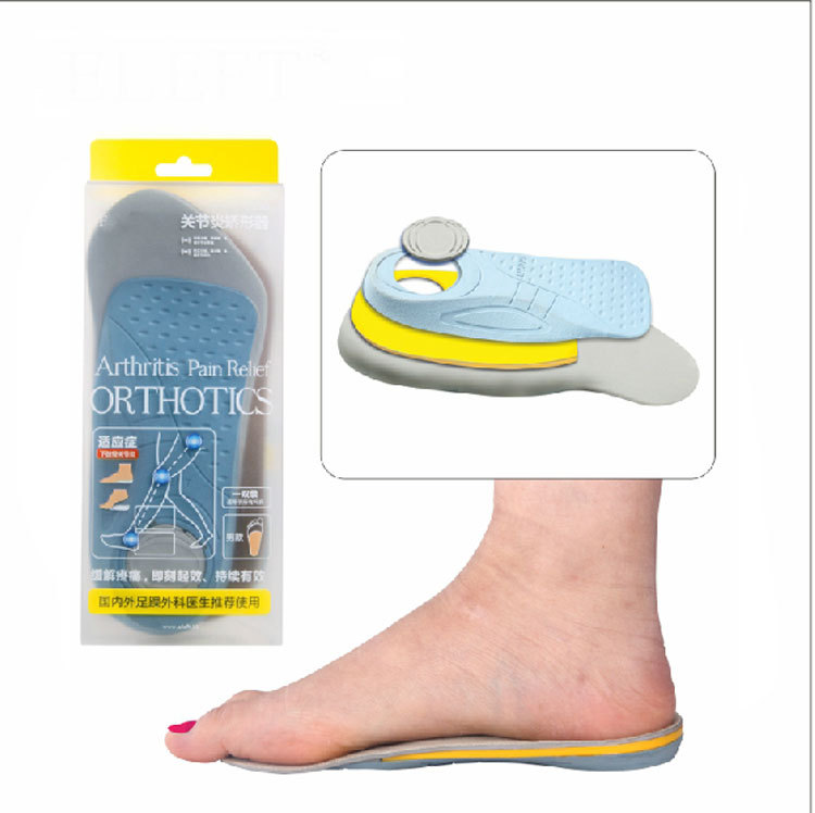
There are several things that you can do to prevent and treat arch pain. This includes Avoiding high heeled shoes, Stretching the calf muscles regularly, Wearing well fitted, comfortable shoes, Using customisedorthotic devices or shoe inserts, Elevating the feet and applying ice and taking over-the-counter anti-inflammatory medications. You can also care for your feet by paying attention to any changes in your feet as you get older. It is normal for feet to lose some of their fat pads as a person ages. Your feet may get bigger, both wider and longer as well. Make sure that you wear shoes that are sturdy, but comfortable, and have your feet measured before you buy shoes to make sure that you are still wearing the right size. Shoe sizes vary from one brand to the next, so it is a good idea to have your feet measured every time you purchase shoes. When choosing shoes, match the shoe to the activity for which it will be worn. Within the broader grouping of athletic shoes, there are different categories with different features. For example, a running shoe has different features than a walking shoe. You may develop some arthritic changes in your feet over time, too. If you notice that you are experiencing more pain in your feet, see your doctor for an evaluation. If the pain is arthritis-related, your doctor may recommend medication or other treatment to slow the progression of the arthritis.
Plantar fasciitis continues to be widely used for the clinical syndrome of undersurface heel pain. However, the use of 'itis' denotes an inflammatory disorder. This is a misnomer as the pathology is not the result of excessive inflammation. Pathological changes are degenerative (but partially reversible) in nature, probably due to repetitive trauma. The plantar fascia is a thick, fibrous band of connective tissue. Its origin is the medial plantar tubercle of the calcaneum. It runs along the sole of the foot like a fan, being attached at its other end to the base of each of the toes. It is a tough, resilient structure that has a number of critical functions during running and walking. It stabilises the metatarsal joints during impact with the ground. It acts as a shock absorber for the entire leg. It forms the longitudinal arch of the foot and helps to lift the arch to prepare it for the 'take-off' phase of the gait cycle.
Causes
Flat feet are a common condition. The condition is normal in infants and toddlers. Flat feet occur because the tissues holding the joints in the foot together (called tendons) are loose. The tissues tighten and form an arch as children grow older. This will take place by the time the child is 2 or 3 years old. Most people have normal arches by the time they are adults. However, the arch may never form in some people. Aging, injuries, or illness may harm the tendons and cause flat feet to develop in a person who has already formed arches. This type of flat foot may occur only on one side. Rarely, painful flat feet in children may be caused by a condition in which two or more of the bones in the foot grow or fuse together. This condition is called tarsal coalition.
Symptoms
Persistant pain and selling under the ball of the foot and extending towards the toes (most commonly the 2nd). Some swelling may be disable on the top of the foot along with redness. Often a sensation of 'walking on the bones for the foot' will be described, and there is a positive Lachman's test. Often a tear will result in the toes splaying (daylight sign) and clawing.
Diagnosis
Diagnosis of a plantar plate tear can often be challenging due to the complex nature of the anatomy of the foot. Careful history taking and an examination of the area of pain is required to determine the extent and cause of the tear. If necessary, further investigations such as x-rays or diagnostic ultrasound may be ordered by your podiatrist to help evaluate the severity of the problem.
Non Surgical Treatment
Rest and icing the swollen area are a good way to practice in-home treatment for mild foot arch pains. Anti inflammatory medications can also be applied to the area. It is important to reduce your activity to avoid worsening the arch pain. Wearing shoes that fit properly is important as well. Make sure to use shoes that are designed with adequate arch support to prevent over pronating. When running, avoid uneven surfaces, as this may contribute to your foot arch pain. Also, orthopedic pads or other padded heel cups and devices can be inserted into shoes to provide support and prevent more strain to the foot.
Surgical Treatment
Fallen arches may occur with deformities of the foot bones. Tarsal coalition is a congenital condition in which the bones of the foot do not separate from one another during development in the womb. A child with tarsal coalition exhibits a rigid flat foot, which can be painful, notes the patient information website eOrthopod. Surgery may prove necessary to separate the bones. Other foot and ankle conditions that cause fallen arches may also require surgery if noninvasive treatments fail to alleviate pain and restore normal function.
Prevention
There are several things that you can do to prevent and treat arch pain. This includes Avoiding high heeled shoes, Stretching the calf muscles regularly, Wearing well fitted, comfortable shoes, Using customisedorthotic devices or shoe inserts, Elevating the feet and applying ice and taking over-the-counter anti-inflammatory medications. You can also care for your feet by paying attention to any changes in your feet as you get older. It is normal for feet to lose some of their fat pads as a person ages. Your feet may get bigger, both wider and longer as well. Make sure that you wear shoes that are sturdy, but comfortable, and have your feet measured before you buy shoes to make sure that you are still wearing the right size. Shoe sizes vary from one brand to the next, so it is a good idea to have your feet measured every time you purchase shoes. When choosing shoes, match the shoe to the activity for which it will be worn. Within the broader grouping of athletic shoes, there are different categories with different features. For example, a running shoe has different features than a walking shoe. You may develop some arthritic changes in your feet over time, too. If you notice that you are experiencing more pain in your feet, see your doctor for an evaluation. If the pain is arthritis-related, your doctor may recommend medication or other treatment to slow the progression of the arthritis.
Will Adult Aquired Flat Foot Necessitate Surgical Teatments ?
Overview
Adult-Acquired Flat Foot Deformity (AAFFD) is mostly caused by a progressive degeneration of the tendon (tibialis posterior) in which sports ths arch in the foot. Because the actual tendon ages or possibly is exposed to repetitive trauma, it stretches out more than time, your all-natural arch in the foot gets for you to be less pronounced and the foot gradually flattens out. Even though it actually is uncertain why this occurs, the issue sometimes appears equally amongst men and ladies - with an growing frequency together with age. Occasionally, the individual get each and every year a new traumatic form of the situation as a result of a fall from a height or even abnormal landing throughout aerial sports like gymnastics or basketball.

Causes
There tend to be numerous leads to involving acquired adult flatfoot, such as fracture or perhaps dislocation, tendon laceration, tarsal coalition, arthritis, neuroarthropathy, neurologic weakness, along with iatrogenic causes. the most frequent cause associated with acquired adult flatfoot will be posterior tibial tendon dysfunction.
Symptoms
The forms of symptoms in which could indicate Adult-Acquired Flat Foot Deformity include foot pain that will worsens more than time, loss with the arch, abnormal shoe wear (excessive wearing on the inner side of shoe through walking around the inner facet in the foot) as well as an awkward look in the foot along with ankle (when viewed from behind, heel and toes apparently go out towards the side). The idea is very important that people help people recognize the particular early the indicators of this condition, as you will find many treatment options, based upon the particular severity, the actual ages of the particular patient, and in addition the desired exercise levels.
Diagnosis
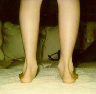
Clinicians must recognize the actual early stage associated with this syndrome which include pain, swelling, tendonitis and disability. The Particular musculoskeletal part with the clinical exam can help determine happens with the disease. The idea is extremely important in order to palpate the particular posterior tibial tendon and also test its muscle strength. This is tested simply by asking affected person to always be able to plantarflex and also invert the actual foot. Joint selection of motion can be should be assessed as well. Stiffness in the joints might indicate longstanding disease causing a new rigid deformity. The weightbearing examination needs in order to be performed as well. Any complete deficiency of your medial longitudinal arch is frequently seen. in later levels your head of the talus bone tasks outward towards the point of your significant "lump" in the arch. Observing your patient's feet through behind shows a substantial valgus rotation with the heel. Coming From behind, the actual "too many toes" sign might be seen as well. This could be when there's abducution of the forefoot within the transverse plane allowing the toes to be seen through behind. Dysfunction in the posterior tibial tendon could be assessed by asking your individual for you to get up on his/her toes around the affected foot. In case they may well be unable to, this indicates the condition will be in the heightened stage with the tendon potentially totally ruptured.
Non surgical Treatment
The following is a introduction to conservative treating acquired flatfoot. Stage 1, NSAIDs as well as short-leg walking cast or walker boot with regard to 6-8 weeks; full-length semirigid custom molded orthosis, physical therapy. Stage 2, UCBL orthosis or even brief articulated ankle orthosis. Stage 3, Molded AFO, double-upright brace, or perhaps patellar tendon-bearing brace. Stage 4, Molded AFO, double-upright brace, or perhaps patellar tendon-bearing brace.

Surgical Treatment
Surgery ought to simply be done in the huge event the pain does not progress following a couple associated with months involving conservative treatment. The Actual kind involving surgery depends around the stage in the PTTD disease. This in addition, it dictated by simply exactly where tendonitis is positioned and how much your tendon is actually damaged. Surgical reconstruction may be extremely complex. Several with the common surgeries include. Tenosynovectomy, taking out the inflamed tendon sheath around the PTT. Tendon Transfer, to be able to augment the particular perform in the diseased posterior tibial tendon with a neighbouring tendon. Calcaneo-osteotomy, sometimes the actual heel bone wants to become corrected to get an improved heel bone alignment. Fusion in the Joints, if osteoarthritis in the foot features set in, fusion of the joints might end up being necessary.
Adult-Acquired Flat Foot Deformity (AAFFD) is mostly caused by a progressive degeneration of the tendon (tibialis posterior) in which sports ths arch in the foot. Because the actual tendon ages or possibly is exposed to repetitive trauma, it stretches out more than time, your all-natural arch in the foot gets for you to be less pronounced and the foot gradually flattens out. Even though it actually is uncertain why this occurs, the issue sometimes appears equally amongst men and ladies - with an growing frequency together with age. Occasionally, the individual get each and every year a new traumatic form of the situation as a result of a fall from a height or even abnormal landing throughout aerial sports like gymnastics or basketball.
Causes
There tend to be numerous leads to involving acquired adult flatfoot, such as fracture or perhaps dislocation, tendon laceration, tarsal coalition, arthritis, neuroarthropathy, neurologic weakness, along with iatrogenic causes. the most frequent cause associated with acquired adult flatfoot will be posterior tibial tendon dysfunction.
Symptoms
The forms of symptoms in which could indicate Adult-Acquired Flat Foot Deformity include foot pain that will worsens more than time, loss with the arch, abnormal shoe wear (excessive wearing on the inner side of shoe through walking around the inner facet in the foot) as well as an awkward look in the foot along with ankle (when viewed from behind, heel and toes apparently go out towards the side). The idea is very important that people help people recognize the particular early the indicators of this condition, as you will find many treatment options, based upon the particular severity, the actual ages of the particular patient, and in addition the desired exercise levels.
Diagnosis
Clinicians must recognize the actual early stage associated with this syndrome which include pain, swelling, tendonitis and disability. The Particular musculoskeletal part with the clinical exam can help determine happens with the disease. The idea is extremely important in order to palpate the particular posterior tibial tendon and also test its muscle strength. This is tested simply by asking affected person to always be able to plantarflex and also invert the actual foot. Joint selection of motion can be should be assessed as well. Stiffness in the joints might indicate longstanding disease causing a new rigid deformity. The weightbearing examination needs in order to be performed as well. Any complete deficiency of your medial longitudinal arch is frequently seen. in later levels your head of the talus bone tasks outward towards the point of your significant "lump" in the arch. Observing your patient's feet through behind shows a substantial valgus rotation with the heel. Coming From behind, the actual "too many toes" sign might be seen as well. This could be when there's abducution of the forefoot within the transverse plane allowing the toes to be seen through behind. Dysfunction in the posterior tibial tendon could be assessed by asking your individual for you to get up on his/her toes around the affected foot. In case they may well be unable to, this indicates the condition will be in the heightened stage with the tendon potentially totally ruptured.
Non surgical Treatment
The following is a introduction to conservative treating acquired flatfoot. Stage 1, NSAIDs as well as short-leg walking cast or walker boot with regard to 6-8 weeks; full-length semirigid custom molded orthosis, physical therapy. Stage 2, UCBL orthosis or even brief articulated ankle orthosis. Stage 3, Molded AFO, double-upright brace, or perhaps patellar tendon-bearing brace. Stage 4, Molded AFO, double-upright brace, or perhaps patellar tendon-bearing brace.
Surgical Treatment
Surgery ought to simply be done in the huge event the pain does not progress following a couple associated with months involving conservative treatment. The Actual kind involving surgery depends around the stage in the PTTD disease. This in addition, it dictated by simply exactly where tendonitis is positioned and how much your tendon is actually damaged. Surgical reconstruction may be extremely complex. Several with the common surgeries include. Tenosynovectomy, taking out the inflamed tendon sheath around the PTT. Tendon Transfer, to be able to augment the particular perform in the diseased posterior tibial tendon with a neighbouring tendon. Calcaneo-osteotomy, sometimes the actual heel bone wants to become corrected to get an improved heel bone alignment. Fusion in the Joints, if osteoarthritis in the foot features set in, fusion of the joints might end up being necessary.
Does Pain In The Arch Need Surgical Procedures ?
Overview
Flexible flatfeet are considered normal in young children because babies are not born with a normal arch. The arch may not form fully until sometime between ages 7 and 10. Even in adulthood, 15% to 25% of people have flexible flatfeet. Most of these people never develop symptoms. In many adults who have had flexible flatfeet since childhood, the missing arch is an inherited condition related to a general looseness of ligaments. These people usually have extremely flexible, very mobile joints throughout the body, not only in the feet. Flatfeet also can develop during adulthood. Causes include joint disease, such as rheumatoid arthritis, and disorders of nerve function (neuropathy).

Causes
The cause of this condition is too much pressure exerted on the arches, and although common in athletes, the condition can happen because you went hiking or climbing, you were lifting heavy objects, or you simply walked too far too vigorously. Pregnancy places extra strain on the arches because of both the additional body weight and the effect of hormones on muscles and ligaments.
Symptoms
Flat feet don't usually cause problems, but they can put a strain on your muscles and ligaments (ligaments link two bones together at a joint). This may cause pain in your legs when you walk. If you have flat feet, you may experience pain in any of the following areas, the inside of your ankle, the arch of your foot, the outer side of your foot, the calf, the knee, hip or back, Some people with flat feet find that their weight is distributed unevenly, particularly if their foot rolls inwards too much (overpronates). If your foot overpronates, your shoes are likely to wear out quickly. Overpronation can also damage your ankle joint and Achilles tendon (the large tendon at the back of your ankle). See your GP if you or your child has flat feet and your feet are painful, even when wearing supportive, well-fitting shoes, shoes wear out very quickly, feet appear to be getting flatter, feet are weak, numb or stiff, Your GP may refer you to a podiatrist (foot specialist).
Diagnosis
Magnetic Resonance Imaging (MRI) can show tendon injury and inflammation but cannot be relied on with 100% accuracy and confidence. The technique and skill of the radiologist in properly positioning the foot with the MRI beam are critical in demonstrating the sometimes obscure findings of tendon injury around the ankle. Magnetic Resonance Imaging (MRI) is expensive and is not necessary in most cases to diagnose posterior tibial tendon injury. Ultrasound has also been used in some cases to diagnose tendon injury, but this test again is usually not required to make the initial diagnosis.
Non Surgical Treatment
Initial treatment of the tear will focus on symptom relief. This may involve the use of anti-inflammatory medications, strapping of the toe, off-loading padding, altered footwear and activity modification. Following this treatment will focus on the underlying causes of the problem, such as flat feet, bunions and hammer toes. Your podiatrist will perform a thorough biomechanical assessment to determine the best course of action to offload the forefoot and decrease the mechanical stress on the area. This injury usually occurs gradually and is best treated in the early stages. Often a customised soft full length orthotic and footwear modifications can prevent the problem from progressing. Occasionally surgery needs to be performed, either frank repair of the plantar plate or in chronic cases a resection osteotomy may be suggested.

Surgical Treatment
Surgical advances have dramatically improved the ability to alleviate the pain and decreased function that millions of Americans experience due to flat feet. Nevertheless, many patients and even some physicians remain unaware of the new procedures, which are best performed by a foot and ankle specialist who has the applicable training and experience.
Stretching Exercises
Start in an L-Sit position. (If you?re hips and hamstrings are tight sit up on a box or phone book to be able to achieve a tall back position. You can even sit on a box with your back supported against a wall!) Keeping the legs straight, but not locked, reach both heels out away from your body to ?Flex? the ankles. Try to avoid pulling back with the toes to flex. Keep the toes relaxed and lead from the heel to hinge the foot into the flexed position. Hold the flexed foot and breathe. Take 3-5 breaths and see if you can reach farther through the heel to deepen the flex on each exhale. To transition to the pointed position, begin by pointing the foot to move the ankles as far as possible in the other direction. Once the ankles have reached their endpoint, use the muscles along the sole of the foot to point the toes. Inhale to continue lengthening out through the top of the foot, exhale to deepen the contraction under the sole of the foot to point the toes. Take 3-5 breaths. Then release the toes, and begin reaching out through the heel to hinge the ankle into the flexed position to repeat the exercise. Continue to flex and the point for 5-10 repetitions.
Flexible flatfeet are considered normal in young children because babies are not born with a normal arch. The arch may not form fully until sometime between ages 7 and 10. Even in adulthood, 15% to 25% of people have flexible flatfeet. Most of these people never develop symptoms. In many adults who have had flexible flatfeet since childhood, the missing arch is an inherited condition related to a general looseness of ligaments. These people usually have extremely flexible, very mobile joints throughout the body, not only in the feet. Flatfeet also can develop during adulthood. Causes include joint disease, such as rheumatoid arthritis, and disorders of nerve function (neuropathy).
Causes
The cause of this condition is too much pressure exerted on the arches, and although common in athletes, the condition can happen because you went hiking or climbing, you were lifting heavy objects, or you simply walked too far too vigorously. Pregnancy places extra strain on the arches because of both the additional body weight and the effect of hormones on muscles and ligaments.
Symptoms
Flat feet don't usually cause problems, but they can put a strain on your muscles and ligaments (ligaments link two bones together at a joint). This may cause pain in your legs when you walk. If you have flat feet, you may experience pain in any of the following areas, the inside of your ankle, the arch of your foot, the outer side of your foot, the calf, the knee, hip or back, Some people with flat feet find that their weight is distributed unevenly, particularly if their foot rolls inwards too much (overpronates). If your foot overpronates, your shoes are likely to wear out quickly. Overpronation can also damage your ankle joint and Achilles tendon (the large tendon at the back of your ankle). See your GP if you or your child has flat feet and your feet are painful, even when wearing supportive, well-fitting shoes, shoes wear out very quickly, feet appear to be getting flatter, feet are weak, numb or stiff, Your GP may refer you to a podiatrist (foot specialist).
Diagnosis
Magnetic Resonance Imaging (MRI) can show tendon injury and inflammation but cannot be relied on with 100% accuracy and confidence. The technique and skill of the radiologist in properly positioning the foot with the MRI beam are critical in demonstrating the sometimes obscure findings of tendon injury around the ankle. Magnetic Resonance Imaging (MRI) is expensive and is not necessary in most cases to diagnose posterior tibial tendon injury. Ultrasound has also been used in some cases to diagnose tendon injury, but this test again is usually not required to make the initial diagnosis.
Non Surgical Treatment
Initial treatment of the tear will focus on symptom relief. This may involve the use of anti-inflammatory medications, strapping of the toe, off-loading padding, altered footwear and activity modification. Following this treatment will focus on the underlying causes of the problem, such as flat feet, bunions and hammer toes. Your podiatrist will perform a thorough biomechanical assessment to determine the best course of action to offload the forefoot and decrease the mechanical stress on the area. This injury usually occurs gradually and is best treated in the early stages. Often a customised soft full length orthotic and footwear modifications can prevent the problem from progressing. Occasionally surgery needs to be performed, either frank repair of the plantar plate or in chronic cases a resection osteotomy may be suggested.
Surgical Treatment
Surgical advances have dramatically improved the ability to alleviate the pain and decreased function that millions of Americans experience due to flat feet. Nevertheless, many patients and even some physicians remain unaware of the new procedures, which are best performed by a foot and ankle specialist who has the applicable training and experience.
Stretching Exercises
Start in an L-Sit position. (If you?re hips and hamstrings are tight sit up on a box or phone book to be able to achieve a tall back position. You can even sit on a box with your back supported against a wall!) Keeping the legs straight, but not locked, reach both heels out away from your body to ?Flex? the ankles. Try to avoid pulling back with the toes to flex. Keep the toes relaxed and lead from the heel to hinge the foot into the flexed position. Hold the flexed foot and breathe. Take 3-5 breaths and see if you can reach farther through the heel to deepen the flex on each exhale. To transition to the pointed position, begin by pointing the foot to move the ankles as far as possible in the other direction. Once the ankles have reached their endpoint, use the muscles along the sole of the foot to point the toes. Inhale to continue lengthening out through the top of the foot, exhale to deepen the contraction under the sole of the foot to point the toes. Take 3-5 breaths. Then release the toes, and begin reaching out through the heel to hinge the ankle into the flexed position to repeat the exercise. Continue to flex and the point for 5-10 repetitions.
Pain In The Foot's Arch Causes Symptoms And Treatments
Overview
Arch pain is often felt as a burning sensation under the long arch of the foot. There are a number of causes of this, but the most common is plantar fasciitis, but can also be due to the strain of any structure in the arch of the foot.
Causes
Arch and heel pain is usually the result of faulty biomechanics (walking gait abnormalities) that place too much stress on the heel bone and the soft tissues that attach to it. This increased stress causes local inflammation and pain. The most common cause of the stress is a condition where the inside arch of the foot flattens more than it should (often known as "over-pronation"). When the arch of the foot flattens, it also gets longer, causing a stretch on the plantar fascia. In response, the heel becomes inflamed where the plantar fascia attaches.
Symptoms
Most flat feet do not cause pain or other problems. Children may have foot pain, ankle pain, or lower leg pain. They should be evaluated by a health care provider if this occurs. Symptoms in adults may include tired or achy feet after long periods of standing or playing sports.
Diagnosis
The diagnosis of high arch (cavus) foot deformity or Charcot Marie Tooth disease can be made by an orthopedic surgeon in the office. Evaluation includes a thorough history and physical examination as well as imaging studies such as X-rays. The orthopedic surgeon will look at the overall shape, flexibility, and strength of a patient?s foot and ankle to help determine the best treatment. Nerve tests may occasionally need to be performed to help confirm the diagnosis.
Non Surgical Treatment
Stretch the fascia. Prop your toes up against a wall, keeping your arch and heel flat so the toes stretch. Hold for a count of 10. Repeat 10 times three or four times per day. Roll a frozen water bottle under the arch. Stretch first then roll out the arch for 10 minutes; you don?t want to stretch the tendon when it?s ice cold. Freeze a golf ball and massage the fascia. Roll the frozen golf ball under the foot, starting from the front and working your way back. Put good pressure on each spot-the medial, center and lateral positions-for 15 seconds before moving to the next area. Then, roll the ball back and forth over the entire foot. Foam roll all muscles on the body above the plantar. Even tight shoulders can cause the condition, as your arm swing can throw off proper hip alignment and footstrike. Bump your arch. Get a commercial insole with an arch bump to push on the plantar and keep it from flexing-it doesn?t matter if you?re an under or overpronator; the plantar needs to be supported and strengthened, Wear the support in all shoes, if possible.

Surgical Treatment
Although most patients with plantar fasciitis respond to non- surgical treatment, a small percentage of patients may require surgery. If, after several months of non-surgical treatment, you continue to have heel pain, surgery will be considered. Your foot and ankle surgeon will discuss the surgical options with you and determine which approach would be most beneficial. No matter what kind of treatment you undergo for plantar fasciitis, the underlying causes that led to this condition may remain. Therefore, you will need to continue with preventive measures. Wearing supportive shoes, stretching, and using custom orthotic devices are the mainstays of long-term treatment for plantar fasciitis.
Prevention
People who run regularly should replace shoes every six months, more frequently if an avid runner. Avoid running or stepping on uneven surfaces. Try to be careful on rocky terrain or hills with loose gravel. Holes, tree stumps and roots are problems if you are trail running. If you have problems with the lower legs, a dirt road is softer than asphalt, which is softer than concrete. Try to pick a good surface if possible. However, if you're racing, be sure to train on the surface you'll eventually run on. Be careful running too many hills. Running uphill is a great workout, but make sure you gradually build this up to avoid injuries. Be careful when running downhill too fast, which can often lead to more injuries than running uphills. Prevent recurrent injuries. Athletes who have experienced ankle injuries previously may benefit from using a brace or tape to prevent recurrent ankle injuries.
Stretching Exercises
Plantar Fasciitis stretches should always be gentle and pain free, if discomfort occurs with or after stretching decrease the intensity and duration of stretches. Stretches can usually be gradually progressed in intensity and duration over time according to individual tolerance. Plantar Fasciitis Stretch 1. Stretch for the right calf muscle (gastrocnemius) and the arch of the right foot (plantar fascia and muscles under the arches). Take your right heel close to the wall and ball of the foot upright against the wall. Move your hips forwards to the wall. Try to keep your right leg straight. Push down through your right heel to increase the stretch. Maintain for 30 seconds, repeat 2-3 times. Plantar Fasciitis Stretch 2. Stretch for the outside belly of the right calf muscle and the arch of the right foot. Take your right heel close to the wall. Turn the ball of your right foot outwards to 2 o?clock position upright against the wall. Move your hips forwards to the wall. Turn your trunk in the opposite direction (i.e. to the left). Try to keep your right leg straight. Push down through your right heel to increase the stretch. Maintain for 30 seconds, repeat 2-3 times. Plantar Fasciitis Stretch 3. Stretch for the inside belly of the right calf muscle and the arch of the right foot. Take your right heel close to the wall. Turn the ball of your right foot inwards to 10 o?clock position upright against the wall. Move your hips forwards to the wall. Turn your trunk in the opposite direction (i.e. to the right). Try to keep your right leg straight. Push down through your right heel to increase the stretch. Maintain for 30 seconds, repeat 2-3 times. Plantar Fasciitis Stretch 4. Stretch for the right achilles tendon and the arch of the right foot. Take your right heel close to the wall and ball of the foot upright against the wall (as for stretch 1). Move your hips forwards to the wall. Bend your right knee forwards into the wall keeping the ball of your foot upright against the wall. Push down through your right heel to increase the stretch. Maintain for 30 seconds, repeat 2-3 times.
Arch pain is often felt as a burning sensation under the long arch of the foot. There are a number of causes of this, but the most common is plantar fasciitis, but can also be due to the strain of any structure in the arch of the foot.
Causes
Arch and heel pain is usually the result of faulty biomechanics (walking gait abnormalities) that place too much stress on the heel bone and the soft tissues that attach to it. This increased stress causes local inflammation and pain. The most common cause of the stress is a condition where the inside arch of the foot flattens more than it should (often known as "over-pronation"). When the arch of the foot flattens, it also gets longer, causing a stretch on the plantar fascia. In response, the heel becomes inflamed where the plantar fascia attaches.
Symptoms
Most flat feet do not cause pain or other problems. Children may have foot pain, ankle pain, or lower leg pain. They should be evaluated by a health care provider if this occurs. Symptoms in adults may include tired or achy feet after long periods of standing or playing sports.
Diagnosis
The diagnosis of high arch (cavus) foot deformity or Charcot Marie Tooth disease can be made by an orthopedic surgeon in the office. Evaluation includes a thorough history and physical examination as well as imaging studies such as X-rays. The orthopedic surgeon will look at the overall shape, flexibility, and strength of a patient?s foot and ankle to help determine the best treatment. Nerve tests may occasionally need to be performed to help confirm the diagnosis.
Non Surgical Treatment
Stretch the fascia. Prop your toes up against a wall, keeping your arch and heel flat so the toes stretch. Hold for a count of 10. Repeat 10 times three or four times per day. Roll a frozen water bottle under the arch. Stretch first then roll out the arch for 10 minutes; you don?t want to stretch the tendon when it?s ice cold. Freeze a golf ball and massage the fascia. Roll the frozen golf ball under the foot, starting from the front and working your way back. Put good pressure on each spot-the medial, center and lateral positions-for 15 seconds before moving to the next area. Then, roll the ball back and forth over the entire foot. Foam roll all muscles on the body above the plantar. Even tight shoulders can cause the condition, as your arm swing can throw off proper hip alignment and footstrike. Bump your arch. Get a commercial insole with an arch bump to push on the plantar and keep it from flexing-it doesn?t matter if you?re an under or overpronator; the plantar needs to be supported and strengthened, Wear the support in all shoes, if possible.
Surgical Treatment
Although most patients with plantar fasciitis respond to non- surgical treatment, a small percentage of patients may require surgery. If, after several months of non-surgical treatment, you continue to have heel pain, surgery will be considered. Your foot and ankle surgeon will discuss the surgical options with you and determine which approach would be most beneficial. No matter what kind of treatment you undergo for plantar fasciitis, the underlying causes that led to this condition may remain. Therefore, you will need to continue with preventive measures. Wearing supportive shoes, stretching, and using custom orthotic devices are the mainstays of long-term treatment for plantar fasciitis.
Prevention
People who run regularly should replace shoes every six months, more frequently if an avid runner. Avoid running or stepping on uneven surfaces. Try to be careful on rocky terrain or hills with loose gravel. Holes, tree stumps and roots are problems if you are trail running. If you have problems with the lower legs, a dirt road is softer than asphalt, which is softer than concrete. Try to pick a good surface if possible. However, if you're racing, be sure to train on the surface you'll eventually run on. Be careful running too many hills. Running uphill is a great workout, but make sure you gradually build this up to avoid injuries. Be careful when running downhill too fast, which can often lead to more injuries than running uphills. Prevent recurrent injuries. Athletes who have experienced ankle injuries previously may benefit from using a brace or tape to prevent recurrent ankle injuries.
Stretching Exercises
Plantar Fasciitis stretches should always be gentle and pain free, if discomfort occurs with or after stretching decrease the intensity and duration of stretches. Stretches can usually be gradually progressed in intensity and duration over time according to individual tolerance. Plantar Fasciitis Stretch 1. Stretch for the right calf muscle (gastrocnemius) and the arch of the right foot (plantar fascia and muscles under the arches). Take your right heel close to the wall and ball of the foot upright against the wall. Move your hips forwards to the wall. Try to keep your right leg straight. Push down through your right heel to increase the stretch. Maintain for 30 seconds, repeat 2-3 times. Plantar Fasciitis Stretch 2. Stretch for the outside belly of the right calf muscle and the arch of the right foot. Take your right heel close to the wall. Turn the ball of your right foot outwards to 2 o?clock position upright against the wall. Move your hips forwards to the wall. Turn your trunk in the opposite direction (i.e. to the left). Try to keep your right leg straight. Push down through your right heel to increase the stretch. Maintain for 30 seconds, repeat 2-3 times. Plantar Fasciitis Stretch 3. Stretch for the inside belly of the right calf muscle and the arch of the right foot. Take your right heel close to the wall. Turn the ball of your right foot inwards to 10 o?clock position upright against the wall. Move your hips forwards to the wall. Turn your trunk in the opposite direction (i.e. to the right). Try to keep your right leg straight. Push down through your right heel to increase the stretch. Maintain for 30 seconds, repeat 2-3 times. Plantar Fasciitis Stretch 4. Stretch for the right achilles tendon and the arch of the right foot. Take your right heel close to the wall and ball of the foot upright against the wall (as for stretch 1). Move your hips forwards to the wall. Bend your right knee forwards into the wall keeping the ball of your foot upright against the wall. Push down through your right heel to increase the stretch. Maintain for 30 seconds, repeat 2-3 times.